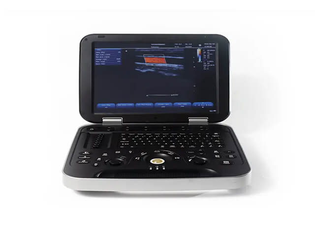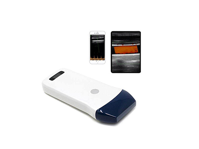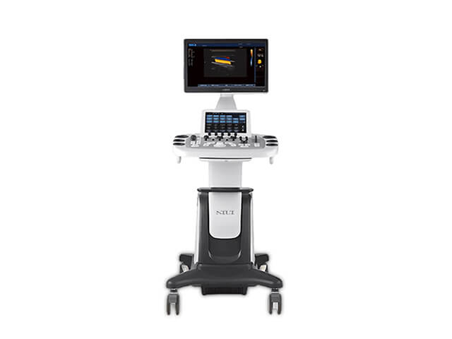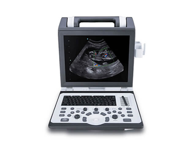
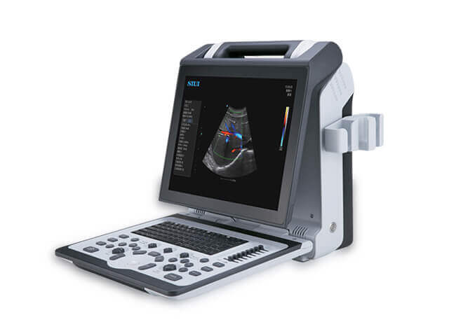
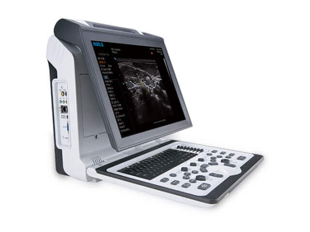
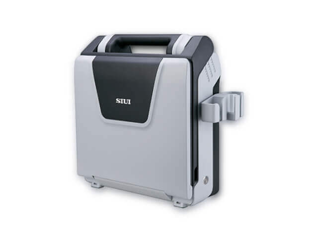


Apogee 2300
Specific for cardiac, pediatric and urologic exam
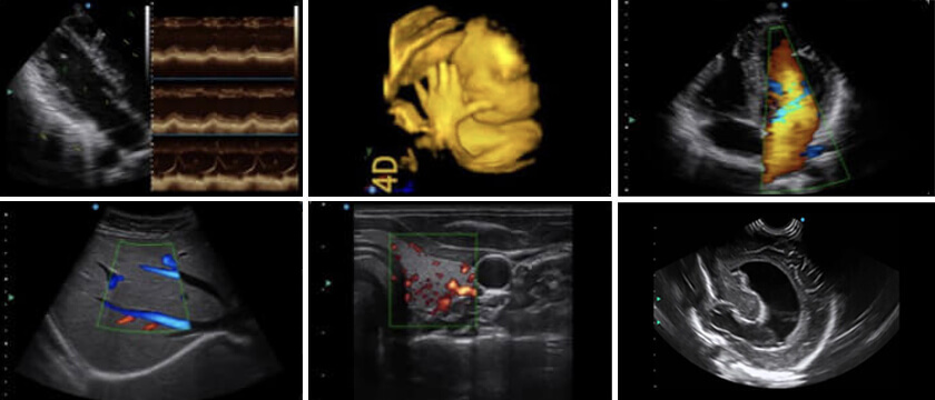
Supports quick cardiac exams with high clinical value: CW, TDI, Auto EF, Auto SG
Comprehensive pediatric assessments covering abdomen, brain and heart, supported by high performance micro convex probe Two types of Bi-plane probes ensure precise diagnosis
Biopsy needle guides increase puncture precision
Residual urine volume calculation and prostate quality evaluation available
The Siui Apogee 2300 is powerful enough to deliver exceptional image quality, but portable enough (9kg) to be quickly and easily transported. it ticked all the boxes for a high performance veterinary ultrasound machine. Other makes and models had come close over the years, but when it came to echocardiography, they always let us down – and usually, it was the quality of the Doppler, both colour flow and spectral.
The Apogee 2300 matches or betters machines such as the Mindray M7, Sonoscape S9, MyLab 30, and even the Philips CX-50 , on both price and performance.
The Siui Apogee 2300 is an ideal solution for private cardiology clinics or small animal vet practices looking to advance their echocardiography capabilities. Its cardiac presets
deliver crystal clear image quality with superior endocardial definition, and tracking to calculate ejection fractions a breeze. For more advanced users, Siui’s Auto-EF software
is also an available add-on, which utilises advances in speckle tracking technology to automatically track the endocardium throughout the cardiac cycle.
Colour flow Doppler can often be a drawback for portable ultrasound machines. They often lack the frame rates required to adequately demonstrate regurgitant flow, particularly
at fast heart rates which tend to go hand-in-hand with common diseases such as mitral regurgitation secondary to degenerative mitral valve disease in dogs. The Apogee 2300
handles this challenge exceptionally well.

★ MFI
+By reducing signal distortion and eliminating unwanted noises, MFI renders premium images with outstanding resolution high contrast and enhanced penetration.
★ XBeam
+The technology helps to ease echo artifacts and improve spatial resolution.
★ Nanoview
+By reducing noises and artifacts, Nanoview is able to present tiny lesions in soften images with distinct tissues and enhanced edge helping to offer reliable diagnostic results.
★ Fusion THI
+Real-time fusing the information from different frequency bands, Fusion THI implements the broadband transmission and reception of harmonic waves.
★ Compact and flexibility
+15-inch LCD monitor with tilting angle, user-orientated control panel, dual probe connectors, replaceable Li-ion Batteries.

1. Lung scanning applications
+Recommend a phased array or convex probe. Lung ultrasound can be performed with a linear probe, but you will be limited to assessing only the top layers (you will not see much beyond the pleura)
2. Rule out heart failure
+ Recommend phased array (as an alternative cause of pulmonary oedema or pleural effusions)
3. Abdominal applications
+ Recommend the convex probe
4. 4.5-7.5MHz broadband microconvex transducer for small animal abdominal work
5. 1.7-4 MHz broadband phased array and/or 4-7 MHz phased array transducers.
6. Continuous Wave Doppler enabled – essential for estimating pulmonary pressures and stenotic valves
7. Tissue Doppler Imaging (TDI) – for a complete assessment of diastolic function
8. Colour M-mode – useful for the qualitative assessment of aortic regurgitation (regurgitant jet in comparison with LVOT width), diastolic function, and cardiac timings, particularly where ECG gating is not available or practical

 Russian
 Español
